Patient L.T.T.R. (69 years old, residing in Tam Ky City, Quang Nam Province) was hospitalized with a cough that lasted for more than 3 months. Recently, coughs have increased with fever and chest pain. Previously, the patient was treated at a lower-level medical facility but suspected that the condition was serious, so he was transferred to Da Nang Hospital.
chest X-ray results recorded images of specific mass damage on the left lung. The chest CT scan showed an image of the accommodation collar with a partial bronchial expansion on the left lung/an image of a deep optic barrier in the tricuspid of the upper - lower left bronchial collar.
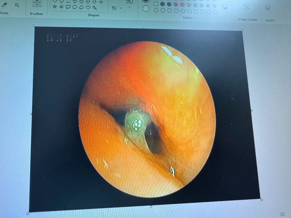
The patient was prescribed a flexible bronchial endoscopy for anesthesia.Doctors successfully removed a piece of bone about 2x1.5cm in size, side-shaped, causing prolonged inflammation.After the procedure, the patient was conscious, had a significant reduction in cough and chest pain, and had good ventilation of the lungs and would be discharged in the near future.
Dr. Hoang Thi Tam, Deputy Head of the Department of Respiratory - Allergy, Da Nang Hospital, said that foreign bodies in the airways are a dangerous cause that can be missed if not thought about and diagnosed early. Foreign objects not only cause chronic pneumonia, coughing up blood, but can also lead to respiratory failure, threatening life.
People need to be especially vigilant when there are symptoms of prolonged cough, recurrent pneumonia, especially after being choked, itchy or eating carelessly. In many cases, foreign objects can enter deep into the airway after a strong cough that the patient does not immediately recognize. bronchial endoscopy is an important technique to detect and remove foreign objects from the airway safely and effectively if any.