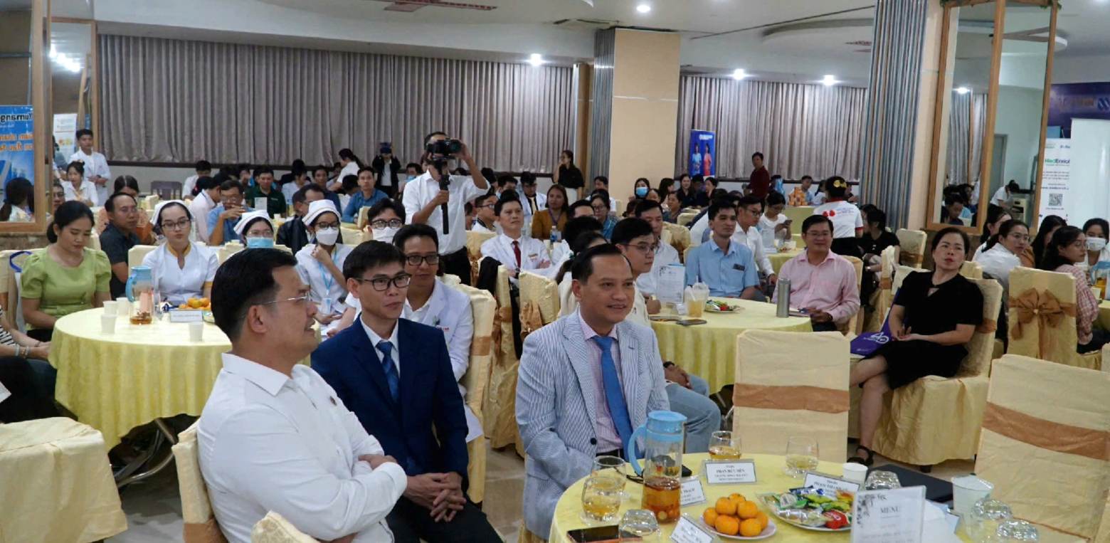
On the afternoon of November 8, Thanh Vu Medic Bac Lieu General Hospital organized a scientific workshop on "Comprehensive update of diagnosis, treatment and the role of diagnostic imaging of liver disease - From metabolic disorders to liver cell cancer".
Doctor Pham Thanh Vu emphasized: liver disease, especially cancer and metabolic disorders, is a big challenge in the Mekong Delta region. The workshop is an opportunity for doctors to exchange professional experiences, update advanced techniques and master modern technologies in diagnostic imaging, helping to detect damage from an early stage, improve treatment accuracy, and serve patients better.
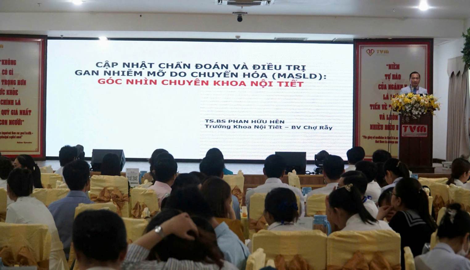
Dr. Phan Huu Hen - Head of the Endocrinology Department of Cho Ray Hospital - updated the latest instructions and emphasized the importance of early identification and intervention of this silent disease to prevent the risk of liver fibrosis and liver cancer. The update of guideline helps the medical team better understand the central role of lifestyle and nutritional adjustment in addition to some newly prescribed drugs to slow down the disease process, protecting the health of high-risk people in the area.
Speakers recommend: Factors such as aflatoxin exposure, alcoholism, smoking and metabolic disorders also contribute to the risk of disease. Periodic liver ultrasound screening combined with biometric markers (AFP, AFP-L3, PIVKA II) helps detect HCC early, especially in high-risk groups. Diagnosis based on dynamic images (CT, MRI) and in-depth tests helps accurately identify damage. Current HCC treatment includes liver surgery, on-site tumor destruction, chemical vasoconstriction, targeted treatment, immunotherapy, liver transplantation and gentle care depending on the stage of the disease.
Doctor Pham Thanh Vu - Chairman of the Board of Directors of Thanh Vu Medic Bac Lieu General Hospital - updated the topic "The role of diagnostic imaging in Gastroesophageal cancer" emphasizing the importance of modern diagnostic technology in early detection and treatment of liver disease. Advanced devices integrated with artificial intelligence (AI) such as MRI 3.0 Tesla and CT Scanner 128 and 160 slices help create a detailed, sharp image, supporting early detection of small tumors and accurately determining the location of damage. In particular, with its superior resolution and sophisticated soft tissue surveying capabilities, MRI 3.0 Tesla can detect early damage to liver, brain and whole body cancer, in many cases effectively replacing PET-CT, reducing costs, not using ionized radiation and being safer for patients.