By mastering neuroendoscopic techniques in a very narrow space of a few millimeters, the Vinmec medical team removed the hematoma completely after only 2 hours of emergency surgery, helping the patient recover miraculously after 5 days.
Signs of old age "hide" dangerous brain damage
Patient Doan Thi Xuan (73 years old, Thanh Hoa) has a history of high blood pressure for many years. 2 days before being admitted to the hospital, she went to the market but forgot her way home, and the family had to look for her. This manifestation was considered by the family to be a sign of memory decline common in the elderly, so they did not take her for examination.
In the early morning of the hospital admission day, she suddenly became drowsy, reacted slowly, sometimes did not recognize relatives, accompanied by severe headaches and nausea. The family immediately took her to Vinmec Times City International General Hospital for emergency treatment in a state of near coma.
Through examination, doctors recorded a noticeable decrease in the patient's consciousness. Suspecting that the patient had intracranial injury, the doctor prescribed emergency magnetic resonance imaging. MRI results showed that the subdural subdural hematoma in the patient's right cerebral hemisphere was large in size, causing serious compression of brain parenchyma. This is the direct cause of conscious disorder in Ms. Xuan.
Extracting more medical history, the doctor was informed by the family that about 3 months ago, she had fallen hard and hit her head but hid it from her children and grandchildren. "This may be the cause of the hematoma in the patient's brain. For the elderly, the blood vessels are atherosclerotic, the walls of the vessels are thin and easily damaged. A seemingly minor injury can cause tearing of the connective veins, forming subdural hematoma and silently progressing into a large blood clot," said BSCKII Nguyen Dinh Huong - Neurosurgeon, Vinmec Times City.
According to Dr. Huong, if not detected and intervened in time, the hematoma will continue to increase in volume, increasing intracranial pressure, causing cerebral edema and irreversible perineal damage. At that time, the patient may fall into a deep coma, hemiplegia, respiratory failure due to brainstem compression, and even death.
Therefore, immediately after the emergency consultation, the Vinmec medical team prescribed emergency surgery to relieve the compression caused by the blood clot and regain the patient's chance of survival.
Double challenges in the operating room and the joy of recovery for patients
With subdural hematoma, usually liquid blood, the doctor can open a small hole in the skull and place a drain to draw the fluid out. However, in this more complex case, the patient's hematoma not only contains liquid, but also blood clots and many internal septum.
“If only traditional drainage is placed, the dense blood and the severed cavities will not be able to drain completely, the risk of residue and recurrence is very high. We decided to perform endoscopic surgery to directly observe inside the subdural cavity, suction all blood clots and remove all mediators,” shared Dr. Huong.
This decision means that the crew must face a double challenge.
The first is to master the technique in an extremely limited space, only a few millimeters, even the smallest movement requires high accuracy. The team performs a very small skull opening to bring a high-resolution endoscopic system into the hematoma cavity, accurately locating the lesion area. Under the magnification image, the surgeon successively suctions out the liquid blood, and at the same time uses specialized instruments to gently break each fibroid wall, removing all remaining blood clots.
The second challenge and also a factor that increases the difficulty of surgery is the risk of damage to atherosclerosis blood vessels in the elderly. Thin vascular walls can cause secondary bleeding even with a small impact, even creating a larger new blood clot than before surgery.
Each step needs to be tightly controlled, meticulously stopped bleeding and ensure that the brain is released and compressed but does not cause further damage," Dr. Huong emphasized.

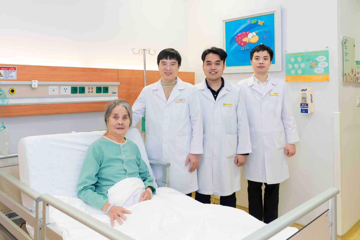
After about 2 hours of surgery, the entire hematoma was removed. Only 30 minutes after surgery, the patient was removed from the endotracheal tube and regained consciousness. 3 days later, MRI images showed no residual hematoma. By the 5th day, Ms. Xuan could walk and live normally.
When I was taken to the hospital, I almost didn't know anything anymore. When I woke up, I saw the doctors next door encouraging and monitoring me very closely. Thanks to timely treatment, now I am recovered and can walk normally, I am really grateful," Ms. Xuan emotionally shared before being discharged from the hospital.
According to BSCKII Nguyen Dinh Huong, endoscopic hematectomy is an effective approach in many complex subdural hematomas. Compared to traditional open surgery, this method helps reduce invasion, maximize the preservation of healthy brain tissue, limit the risk of infection and neurological complications, and shorten recovery time.
The success of the surgery not only opens up recovery opportunities for patients, but also demonstrates the ability to master specialized Neurosurgery techniques. With a modern diagnostic imaging system, emergency consultation procedures and a team of experienced surgeons, Vinmec continues to affirm its strength in treating complex neurological diseases, contributing to improving survival opportunities and quality of life for patients.
To be consulted and examined at Vinmec Times City, please contact to make an appointment through the Vinmec website at https://www.vinmec. com/vie/co-so-y-te/benh-vien-da-khoa-quoc-te-vinmec-times-city-17265-vi-hoi-suc-cap-cuu or download the MyVinmec application.


