Unexpected discovery of skull stenosis from easily overlooked signs
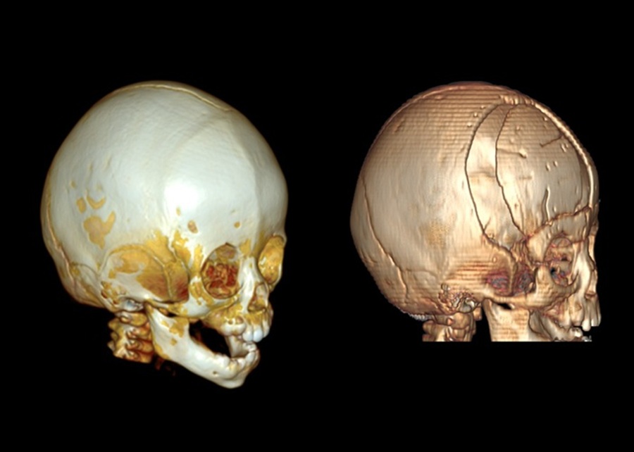
When M.T. was 10 months old, the baby's mother discovered that her child had signs of facial imbalance, slightly misaligned eyebrows, and a deformed skull, so she took the baby to Vinmec Times City for in-depth examination. Through examination and 3D reconstructive brain CT scan, doctors determined that the baby had a skull stenosis due to adhesion to the right parietal frontal joint, causing skull deformities in the forehead and cecumen.
Dr. Dong Pham Cuong - Director of Vinmec Times City Neurocenter - said that skull stenosis is a congenital defect found in young children with a incidence rate of about 1/2,500 children. The main cause of the disease is due to the joint line between the skull bones closing or adhering abnormally early, affecting the normal development of the skull and brain.
Depending on the location of the skull joints that are adhered early, the manifestations of the disease are different, but the common characteristic is that the head and face are unbalanced, with abnormal shapes. Initial symptoms may be vague, easily overlooked if parents do not pay attention to observation, or confuse the phenomenon of distorted head in children due to lying down a lot and will disappear on their own as children grow up.
Untreated skull fractures can cause slow brain development, affect nerve functions, such as language and movement, leaving aesthetic defects that make children insecure and difficult to integrate into society when they grow up. Currently, skull reconstruction surgery is still the optimal treatment for this disease.
Surgery needs to be performed early when the skull is still soft and the brain has not been damaged. If delayed, the skull will become harder, clearly deformed, making the surgery complicated, possibly leaving a large incision and posing many post-operative risks," Dr. Cuong said.
Applying 3D printing technology to plan surgery
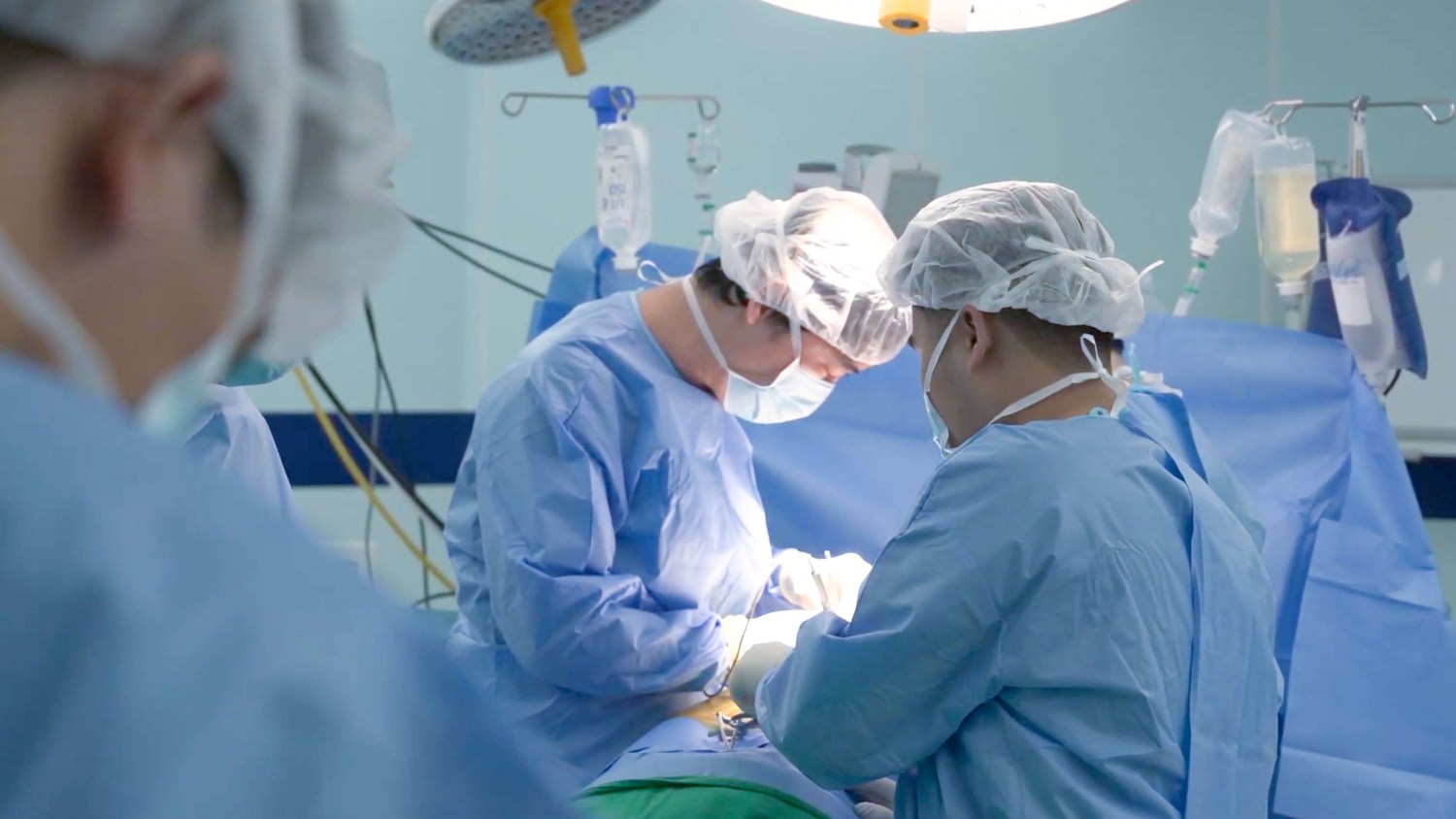
Vinmec Times City doctors consulted and determined that this was a relatively complex surgery because the child was young, while skull reconstruction is a major surgery, causing a lot of blood loss. Therefore, the problem posed for the surgical team is to develop an optimal surgical plan, minimizing blood loss to ensure safety for the child. On the other hand, the child's skull is thin, the nervous system is not yet complete, requiring surgery to achieve absolute accuracy, no mistakes, even the smallest ones.
Faced with these challenges, the surgical team including specialists in neurosurgery and plastic surgery decided to reconstruct the entire skull structure of the patient's child, applying 3D printing technology to create an intuitive skull model, serving the pre-operative "test surgery".
Usually with skull resections, doctors rely on computer-based CT scan data on computers for surgery. However, this method has certain limitations in visualizing the real space and operating procedures.
With a 3D visual skull model, doctors can accurately calculate the surgical path, bone incision path, eyebrow shape direction, placement of splints and fixed screws. This method both ensures aesthetics and avoids the skull affecting brain development, compressing the eye socket, avoiding risks that may occur when children grow up.
The official surgery took place smoothly without any errors. Doctors used self-dissolving bone fixation devices instead of titanium splints to suit the physiological development of children's bones. Self-dissolving bone fixation materials still ensure stability during the bone healing stage, but will completely decompose when the bone structure has stabilized.
Immediately after surgery, baby M.T. was transferred to the intensive care unit for continued monitoring and special care. Vital signs were stable, the baby was awake, and responded well to the treatment regimen. In particular, the skull shape of the patient was significantly improved, and the face, eyebrows, and mouth became more balanced.
According to Dr. Cuong, if well cared for, baby M.T. has a complete opportunity to develop physically normally and return to life like her peers.
The application of artificial intelligence and 3D printing technology is a new solution to help improve accuracy and limit errors in skull structure reconstructive surgeries to treat skull stenosis in children," Dr. Cuong said.
Currently, the Vinmec Health System is the first and only medical unit in Vietnam to apply 3D technology to routine diagnosis and treatment. This technology has been applied by Vinmec in many specialties, such as orthopedic trauma, thoracic surgery... with many complex cases successfully performed, such as reconstruction of thoracic defects with titanium material, pelvic replacement, and complete femur replacement in pediatric patients.
To be consulted and examined with Dr. Dong Pham Cuong, please contact to make an appointment through the Vinmec website at https://www.vinmec. com/vie/chuyen-gia-y-te/dong-pham-cuong or download the MyVinmec application.


