An international scientific project called Human Organ Atlas is revealing the structure of the human body with unprecedented detail, from all internal organs to the structure at the cellular level.
According to the research team, this system can display 3D images of the brain, heart, lungs, liver, kidneys and many other organs with accuracy at a scale of 1 micron, about 50 times thinner than a human hair.
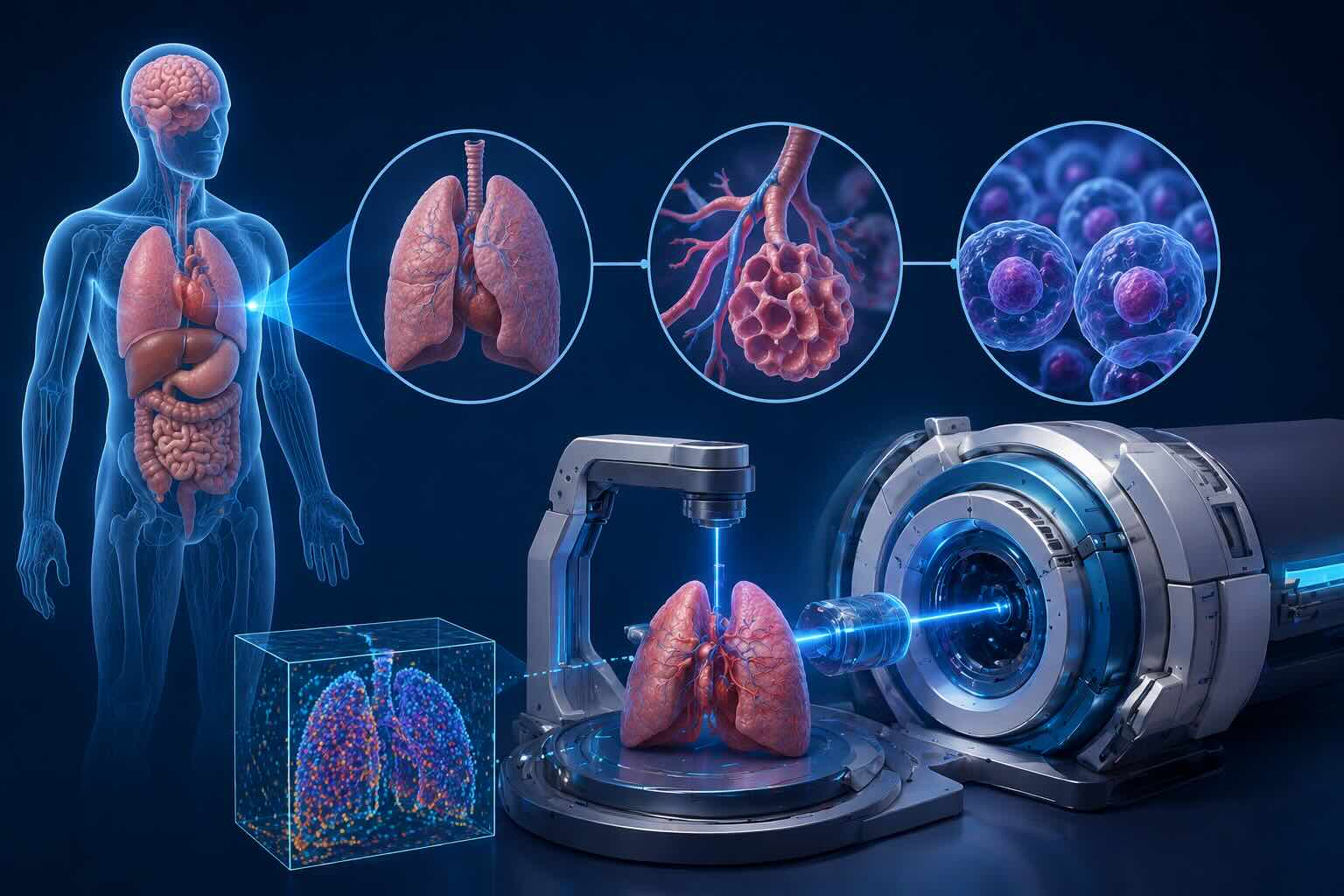
The project uses HiP-CT technology, combined with super-powerful X-rays created from a particle accelerator at the European Synchrotron Radiation Facility.
According to scientists, this X-ray system has a much stronger brightness than conventional X-ray machines in hospitals, allowing observation of organ structures at the cellular level without destroying tissues.
Paul Tafforeau - a scientist in charge of beam systems at the European Synchrotron Radiation Facility said that the project aims to provide open data sources for researchers, doctors and those interested in the structure of the human body.
To date, the project has collected data from 54 donors, creating images of 87 agencies and hundreds of 3D data sets.
These data have helped scientists detect many micro-level pathological signs, including vascular damage in the lungs of patients who died from COVID-19 or characteristics related to endometriosis.


