On July 17, Tu Du Hospital informed that the unit and Children's Hospital 1 had just performed fetal surgery on Ms. P.T.B.T (36 years old, Hiep Chanh Ward, Ho Chi Minh City). This is the 8th successful fetal intervention to give birth to a healthy baby.
Ms. T has been married for 16 years, has never had children and had a miscarriage in 2022. This time, she accidentally discovered that she was pregnant at 8 weeks and 5 days. Ms. T went for a routine pregnancy check-up 6 times, at 12 weeks for low-risk NIPT screening.
At 24 weeks, an ultrasound survey of the figure revealed severe congenital heart defect. After that, Ms. T went to the Cardiovascular Institute for a double check-up and discovered that the fetus had severe narrowing of the pulmonary artery valve, severe 3-leaf valve opening and right hypopharynx.
In April, Ms. T and her husband went to Tu Du Hospital and had a prenatal ultrasound and discovered that the fetus had not had a pulmonary valve opening at 25 weeks and 1 day - the peritoneal wall was closed. Doctors consulted and conducted a liver culture to detect any Chromatic Abnormalities on the CGH Array. The child was closely monitored at Tu Du Hospital and Children's Hospital 1.
In May, when she was 29 weeks and 2 days pregnant, with an estimated weight of 1.4 kg, the team of the 2 hospitals consulted and assessed that the hysterectomy had to progress - hysterectomy had to decrease, with the risk of stillbirth or the risk of not being able to repair the circulation of the 2 hysterects after giving birth. The team decided to open a fetal heart and perform a pulmonary valve after carefully considering the benefits and risks of surgery.
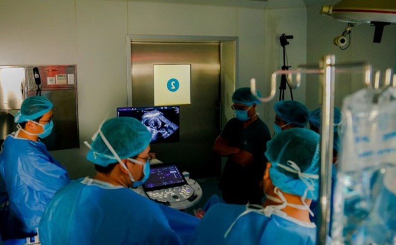
The surgery was performed on the morning of July 15. After 4 hours of intervention surgery, the mother's blood flow was stable, the ultrasound examined the fetal heart and showed that the blood flow was quite good, without any recorded events.
In the days after the intervention, Ms. T felt a tight abdomen and a throbbing appeared. Doctors used the maximum canker slash and lasted 2 weeks, the pregnant woman was stable and discharged from the hospital.
After that, Ms. T had a regular check-up every 2 weeks, and obstetricians assessed the comprehensive development of the fetus with all indicators within the normal range. The right amniotic cavity of the fetus increases in size with each ultrasound with the right amniotic function in both the right atoms and the right atoms, so the accumulation and absorption are good.
In July, when the fetus was 37 weeks 1 day old, with an estimated weight of 3.1 kg, the index on fetal ultrasound was normal. On July 14, Ms. T was consulted by doctors and decided to terminate the pregnancy when she was 38 weeks old.
On July 15, the crew of the two hospitals performed cesarean section for Ms. T. After 5 minutes, a baby boy was born weighing 3.2 kg.
The child's respiratory and circulatory condition was good, the on-site ultrasound examined the baby's heart and found that the 4 heart cells were quite balanced, the blood flow to the pulmonary artery valve was quite stable. The child was transferred to Children's Hospital 1 for further monitoring.
After the first day of surgery, Ms. T was able to walk on her own.