Pregnant woman L.T.H.N (24 years old, An Giang) was transferred to the hospital at 5:39 p.m. on October 1 with severe anemia, pale mucous membranes, pale skin, lethargy, balloon compression via endotracheal tube, rapid and weak pulse, difficult to measure blood pressure, blood clotting disorder, severe metabolic acidosis, using high doses of vasopressors, the patient was giving birth for the third time.
On the same day of admission to the hospital at the upper level, the pregnant woman underwent cesarean section with a history of placenta previa at 31 weeks. However, after surgery, the patient developed severe postpartum hemorrhage, uncontrolled bleeding, uterine atony, and was given emergency treatment, partial hysterectomy, and 18 units of blood and blood products were transfused.
At Can Tho General Hospital, the patient was actively resuscitated, ventilated, anti-acid, continued red blood cell transfusion... When the condition allowed, the patient was transferred for abdominal CT scan with contrast with the diagnosis: Hemorrhagic shock with complications of multiple organ failure, coagulation disorder, post-operative partial hysterectomy due to postpartum hemorrhage, severe metabolic acidosis.

Abdominal CT scan with contrast showed an image of a vascular leak.
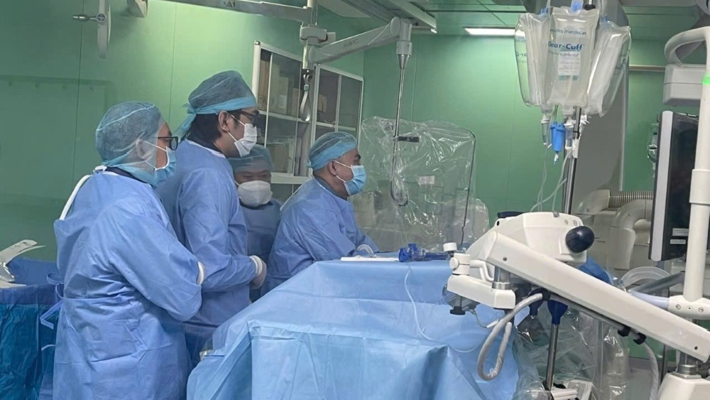
The patient was indicated for digital subtraction angiography and embolization to treat hemostasis of the organs in an emergency. The results showed multiple branches of the internal iliac arteries on both sides, vasospastic lesions with drug leakage in the pelvic area supplied by the left internal iliac branch. The location was determined and embolization was performed with a glue mixture. The examination showed that there was no more leakage. When the patient's vital signs were stable, he was transferred to the Intensive Care - Anti-Poison Department for further monitoring.
At noon on October 2, the patient showed signs of recurrent internal bleeding. Doctors performed a second digital subtraction angiography (DSA) scan and embolization to stop the bleeding of the organs. The results showed an image of the extravasation being supplied with blood from the superior mesenteric artery branch, determining the location for embolization with a glue mixture. The procedure lasted 40 minutes, and the patient was stable.
After 2 days of intervention, the patient's blood loss was controlled. However, due to massive blood loss, the patient developed multiple organ failure, and the doctors of the Intensive Care - Anti-Poison Department continued to perform continuous blood filtration while continuing to transfuse blood and blood products.

During the emergency and resuscitation process at the hospital, the patient was transfused with 52 units of blood and blood products. The child's health is stable and is being cared for at home.


