From silent symptoms to pivotal decision
According to Dr. Doan Duc Dung - Director of the Cardiovascular Center, Vinmec Times City International General Hospital, Mr. T has a history of severe aortic valve disease accompanied by many complex cardiovascular diseases. Five months before being hospitalized, he had a biological aortic valve replacement at a medical facility outside the Vinmec system.
About 2 months after surgery, the patient began to feel tired, short of breath when exerting himself but no severe chest pain appeared - a common sign of acute aortic syndrome. A week before being hospitalized, the condition of shortness of breath increased significantly, causing him to have a re-examination. The results showed that the patient had a type A aortic aneurysm accompanied by heart failure, the rate of hemolysis was only about 30%. Faced with the risk of aortic rupture, he was urgently transferred to Vinmec Times City for emergency surgery.
The biggest challenge is that emergency surgery is performed on patients who have had heart valve replacements before. Having to have surgery again after a short time significantly increases the risk of complications and death during surgery, because the structures in the chest have formed a complex adhesion after the previous intervention," said Dr. Dung.
The surgery was performed by a cardiovascular surgery team in coordination with anesthesia and intensive care. As soon as the chest was opened, doctors encountered many difficulties due to the adhesion tissue around the heart and large blood vessels. After approaching the aorta, the team recorded that the ascending aorta had swollen about 7 cm, the dissection line extended close to the aorta.
When directly assessing the damage at the base of the aorta, surgeons discovered that the dissection line was not only localized in the ascending aorta but also spread down the right coronary sinus, causing severe stenosis of the right coronary artery.
This is a particularly dangerous situation. Usually, after the aortic segment is replaced, the coronary artery openings will be re-plugged into the artificial vessel segment to maintain myocardial irrigation. However, in this case, the right coronary artery opening has been damaged by the dissection process. If it continues to be preserved and re-plugged in the usual way, the risk of angio stenosis or obstruction after surgery is very high, which can lead to acute right ventricle myocardial infarction with a high mortality rate.
Faced with that situation, the team decided to proactively stitch the right coronary opening, and at the same time bridge the aorta - right coronary artery with an inverted venous segment to restore the blood flow to nourish the heart muscle. This solution helps eliminate the risk of right coronary artery blockage after surgery, while protecting the heart muscle from acute infarction complications during and after surgery.
At the same time, doctors replaced the entire severed upper aortic segment with an artificial vascular tube, re-implanted the left coronary artery and preserved the biological aortic valve that had been replaced before because the valve was still working well. Keeping the old valve helps limit the level of intervention, preventing patients from having a second valve replacement.
After more than 2 hours of using the extracorporeal circulation system, the patient's heartbeat returned to stability. The esophageal ultrasound right in the operating room showed that the biological valve functioned well, heart function was preserved, and no significant abnormalities were recorded.
According to Dr. Doan Duc Dung, each case of aortic dissection has its own characteristics of lesions, requiring a personalized treatment strategy. Evaluating lesions right in surgery and choosing appropriate treatment options plays an important role in limiting complications and optimizing treatment outcomes.
Multidisciplinary power in treating complex cardiovascular diseases
After surgery, the patient continues to face many risks of complications due to complex medical background and previous heart failure.
In the early days after surgery, Mr. T's hemodynamics were not stable, at times losing about 800 ml of blood through chest drainage, it was necessary to use vasopressors and blood transfusions to maintain circulation. In parallel with intensive resuscitation, the team of doctors early implemented a nutrition and respiratory rehabilitation program, helping the patient successfully quit the ventilator and improved his physical condition.
Another challenge appeared when the patient had acute atrial fibrillation, causing hypotension and hemodynamic effects. Complications were detected and treated promptly by the resuscitation team, bringing heart rate back to normal, while combining medical treatment to control arrhythmia and prevent recurrence.
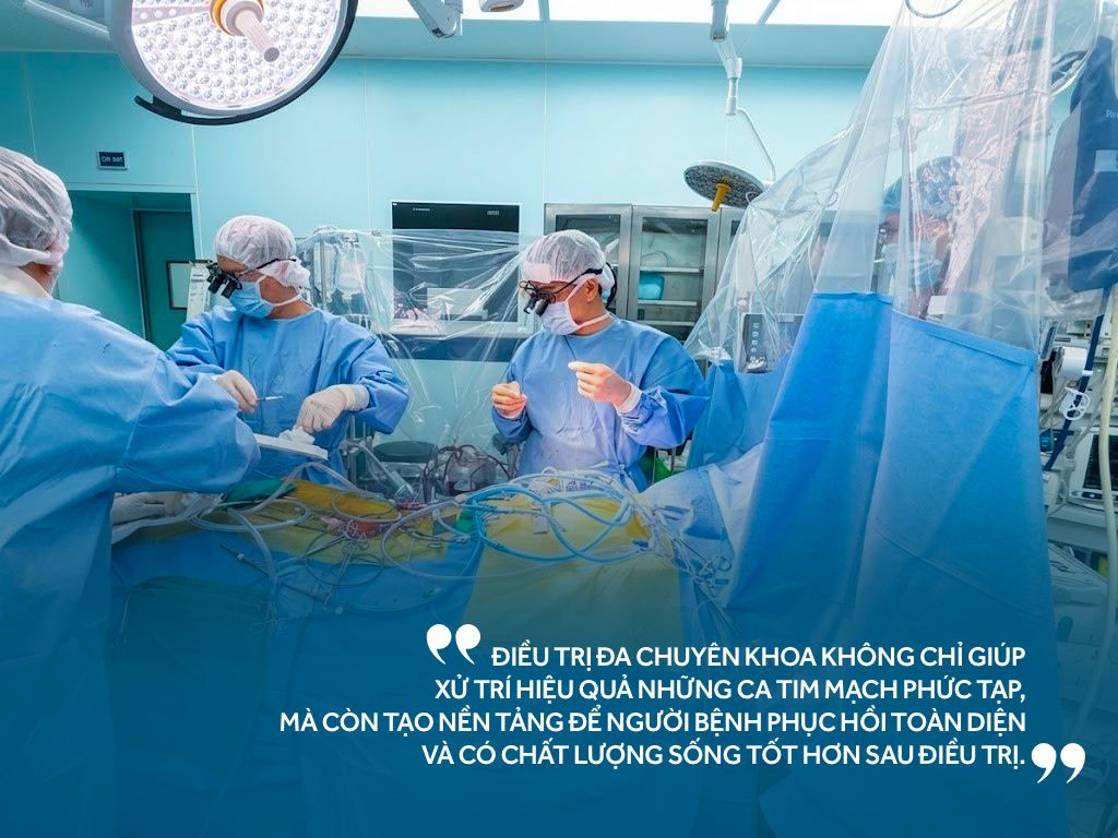
Thanks to the close coordination between cardiology, diagnostic imaging, anesthesia, intensive care, nutrition and functional rehabilitation, the patient recovered well after nearly two weeks of treatment. When discharged from the hospital, Mr. T was alert, his heart rate was stable, the symptoms of shortness of breath were significantly reduced, and the function of left ventricular contraction was significantly improved compared to before surgery.
At Vinmec Times City, treating complex cardiovascular cases is not just an effort of one specialty. Each decision is built on close coordination between many specialties, from diagnosis, surgery, resuscitation to rehabilitation. This comprehensive and individualized approach helps us not only treat acute complex diseases, but also aim for a long-term goal of improving the quality of life for patients," Dr. Doan Duc Dung shared.