Patient D.T.K. (18 months old, Kien Giang province) was admitted to the City Children's Hospital on December 25.
According to the family, while eating peanuts, the child suddenly coughed violently and turned purple. The child's father gave first aid by removing the foreign object, but the child continued to cough violently, turned purple and had difficulty breathing.
After that, he was taken to the district hospital, where he was given first aid by oxygen, and then transferred to the provincial hospital.
At the provincial hospital, the child was given a chest X-ray and diagnosed with suspected foreign body in the airway/congenital heart. After receiving oxygen support, the child was transferred to the City Children's Hospital for further treatment.
Upon admission, the patient showed signs of restlessness, cyanosis, required oxygen, warm extremities, sunken chest, wheezing when inhaling, and hoarseness. Lung auscultation revealed coarse but even lung sounds on both sides, soft abdomen, and non-enlarged liver and spleen.
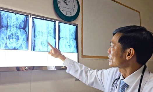
The patient was ordered to have a chest CT scan, the results showed a foreign object in the trachea.
The foreign object was 4x6x11 mm in size (anterior - posterior - horizontal - high), located close to the anterior wall of the trachea, causing near-complete stenosis of the trachea at this location.
Dr. Nguyen Minh Tien - Deputy Director of the City Children's Hospital - said that the patient was diagnosed with a foreign body in the airway with severe respiratory failure complications. Immediately, the doctors provided breathing support, administered antibiotics, and consulted with the respiratory endoscopy team to anesthetize and treat the foreign body.
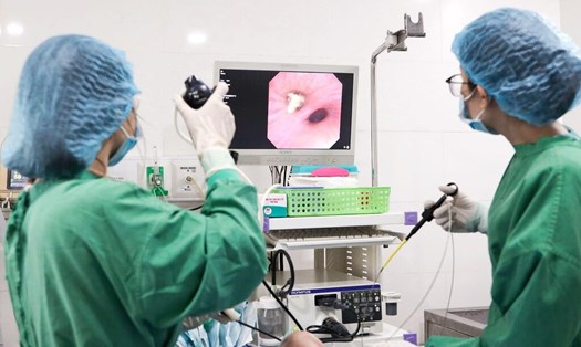
During the endoscopy, doctors used a flexible endoscope through the mouth, recording that the hypopharynx and larynx were clear, and the two vocal cords opened and closed well.
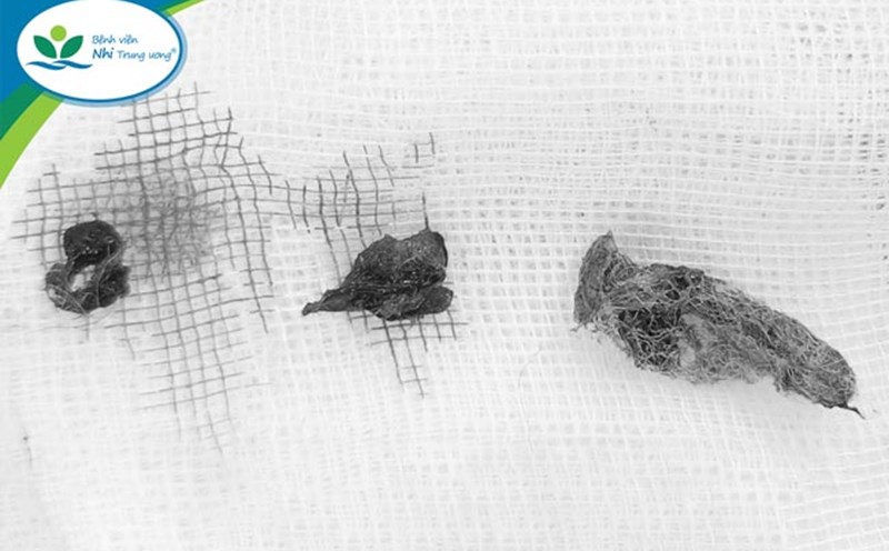
After anesthetizing the vocal cords, the endoscope was continued to be inserted into the trachea, detecting a semicircular foreign body suspected to be a peanut in the middle 1/3 of the trachea, causing almost complete obstruction of the airway. Around the tracheal wall, there were many white pseudomembranes.
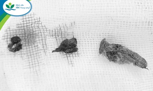
Using forceps to remove the foreign object, the doctors were able to remove many small pieces of the polygon and take out the entire foreign object. After the procedure, the patient was awake and had good communication.


