The rarest ectopic pregnancy
On March 31, Tu Du Hospital said that it had coordinated with Cho Ray Hospital to successfully operate and save Ms. L.T. T (36 years old, Dong Nai) who was carrying an ectopic pregnancy in the abdominal cavity in an extremely rare and dangerous location, directly threatening her life. If detected late, the risk of complications is very high.
Patient T has a history of 2 natural births and had undergone endoscopic surgery to remove the left ear canal due to ectopic pregnancy more than 11 years ago. This pregnancy was unintended. When abdominal pain gradually increased, the patient came to the hospital in Dong Nai and was suspected of being about 10 weeks pregnant in the abdomen, then was urgently transferred to Tu Du Hospital on March 26.
At the Emergency Department, the pregnant woman was examined, consulted and underwent in-depth paraclinical tests. The results determined that there was a live fetus of 10-11 weeks located behind the peritoneum, close to the abdominal aorta, right below the branching position of the left renal artery. The fetal mass showed signs of adhesion to the abdominal aorta wall about 10mm, surrounded by a lot of fluid, suspected of progressive bleeding.
Doctors determined that this was one of the rarest ectopic fetuses, with a risk of large blood vessel rupture causing death if not intervened in time. The hospital activated an inter-hospital red alert, inviting a blood vessel surgical team from Cho Ray Hospital to coordinate treatment. The team consulted and developed an optimal surgical plan to ensure maximum safety for the patient.
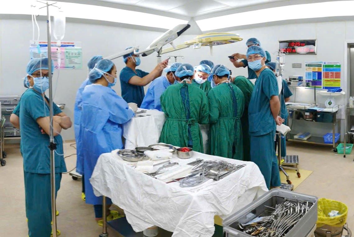
The surgery lasted 3 hours
The surgery was performed at 11:45 PM on the same day. When opening the abdomen, the team recorded a diffuse bleeding in the posterior peritoneum area. The fetal mass was located close to many important structures such as the abdominal aorta, arteries and veins of the left kidney, and left urethra.
Doctors meticulously dissected every millimeter to avoid damage to major blood vessels, successfully preserving the left veins and kidney artery, and at the same time, safely treating the attached fetal mass of the abdominal aorta. The left ureter was also exposed and protected.
The fetal mass is located behind the peritoneum and still carries the left genital artery. During the dissection, this genital artery was partially damaged causing bleeding, the team promptly handled it and patiently restored and preserved it effectively. Finally, the fetal mass and placenta attached to the left kidney area were completely removed.
After nearly 3 hours, the surgery successfully ended with a blood loss of about 500 ml. The patient did not need a blood transfusion.
After 2 days post-operative, Ms. T was completely awake, vital signs were stable, the surgical wound was dry, and she could walk and eat almost normally. By the morning of March 31, Ms. T recovered almost completely and was discharged from the hospital.


