The patient is Mr. Nguyen Van T. (53 years old, residing in Lang Co, Phu Loc district, Hue city), with a history of bronchial asthma, hospitalized with a cough that lasted for more than 5 months, accompanied by frequent diarrhea and chest pain.
According to the story, about 5 months ago, Mr. T. choked on fish bones and had a strong cough during a meal. Due to unclear symptoms, he did not see a doctor. When his cough became more and more serious, he went to a lower-level medical facility and was given a chest CT scan.
The image shows a thin, high foreign object (about 300 HU), about 5mm in size, located in the right mediastinal bronchi, about 32mm from the air duct division, with thick bronchial walls, mucus and fiber bands in the lung parenchyma.
The patient was transferred to Da Nang Hospital for further treatment. After performing paraclinical tests, doctors from the Department of Respiratory - Allergy Immunology prescribed an endoscopy of the sub-menthal bronchium under anesthesia.
Endoscopy results showed that the foreign object was a fish bone beat with three sharp edges, about 1.4 x 1.5cm in size, located in the bronchial median fossa between the median fossa and the fossa under the right lung. The foreign object was safely removed and checked without recording rumors or secondary damage. After the procedure, the patient was alert, the symptoms of cough and chest pain were significantly reduced, the lungs were well ventilated, and were recovering stably.
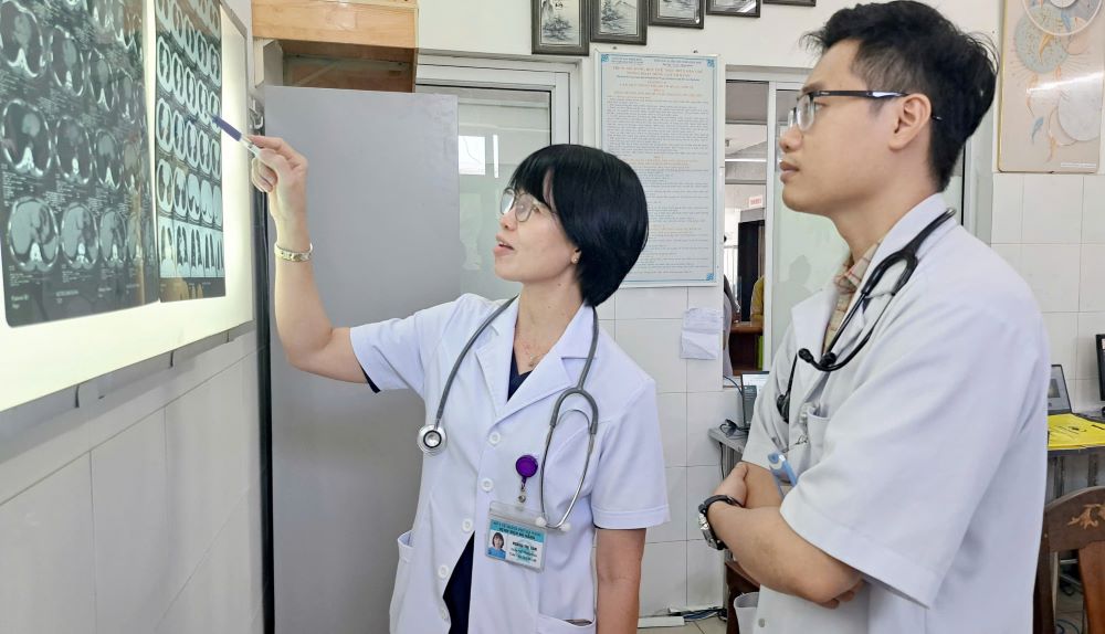
According to Dr. Hoang Thi Tam, Deputy Head of the Department of Respiratory - Allergy, Da Nang Hospital, foreign bodies in the airways in adults are often missed due to atypical symptoms. Many people forget the incident after a bowel movement, only when their cough is prolonged and pneumonia recurs go to the clinic, then the foreign object has existed for too long and causes complications, Dr. Tam shared.